When comparing microscopes, one specification tends to dominate product descriptions:
Magnification.
40×.
400×.
1000×.
1600×.
Even 2000× or 3000×.
It is easy to assume that a microscope with the highest magnification must also provide the most detail.
But this is one of the most common misunderstandings in microscopy.
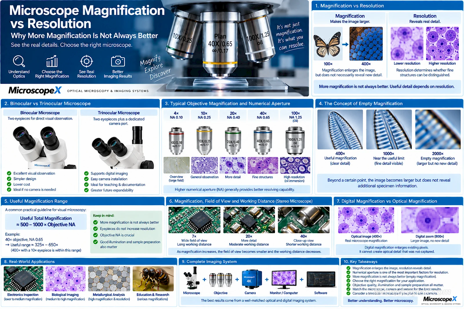
Magnification makes an image larger.
Resolution determines whether additional detail can actually be distinguished.
A microscope can magnify an image dramatically without revealing any new specimen information. When this happens, the result is known as empty magnification.
For most microscopy applications, the better question is therefore not:
How much can this microscope magnify?
It is:
How much useful detail can this microscope resolve?
Understanding the difference between magnification and resolution helps you choose the right microscope, objective lens and camera while avoiding misleading magnification specifications.
This guide explains magnification, numerical aperture, optical resolution, useful magnification, empty magnification and digital magnification in practical terms.
For a general introduction to microscope optics, see Optical Microscope Basics.
Magnification vs Resolution at a Glance
| Property | Magnification | Resolution |
|---|---|---|
| What it describes | How large the image appears | Ability to distinguish fine details |
| Main controlling factors | Objective and eyepiece magnification | Numerical aperture, wavelength and optical quality |
| More is always better? | No | Generally, higher resolving power is desirable |
| Can it reveal new detail? | Only when resolution supports it | Yes |
| Main microscope component | Objective + eyepiece | Primarily objective |
| Common mistake | Buying based on maximum magnification | Ignoring numerical aperture |
| Limitation | Empty magnification | Diffraction and optical system performance |
The distinction between these two concepts is fundamental to microscope selection.
What Is Microscope Magnification?
Magnification describes how much larger an object appears through the microscope compared with viewing it directly.
For a traditional compound microscope, total visual magnification is approximately:
Total Magnification = Objective Magnification × Eyepiece Magnification
For example:
10× objective × 10× eyepiece = 100× total magnification
and:
40× objective × 10× eyepiece = 400× total magnification
A typical biological microscope may use:
- 4× objective
- 10× objective
- 40× objective
- 100× objective
with a 10× eyepiece.
The resulting magnifications are:
| Objective | Eyepiece | Total Magnification |
|---|---|---|
| 4× | 10× | 40× |
| 10× | 10× | 100× |
| 40× | 10× | 400× |
| 100× | 10× | 1000× |
This is why many biological microscopes are described as 40×–1000× systems.
Browse Compound Microscopes for examples of high-power microscope systems.
How Is Stereo Microscope Magnification Calculated?
Stereo microscopes work somewhat differently because many use a continuously adjustable zoom optical body.
A simplified calculation is:
Total Magnification = Zoom Magnification × Eyepiece Magnification × Auxiliary Objective
Suppose a stereo microscope has:
- 0.7×–4.5× zoom body
- 10× eyepieces
- 1× auxiliary objective
The resulting range is:
7×–45×
If you add a 0.5× auxiliary objective:
3.5×–22.5×
If you add a 2× auxiliary objective:
14×–90×
But increasing stereo microscope magnification also changes:
- Field of view
- Working distance
- Depth of field
- Practical usability
This is why the highest magnification is rarely the best setting for every application.
Explore Stereo Microscopes for continuous-zoom systems used in industrial inspection, electronics and laboratory work.
What Is Microscope Resolution?
Resolution describes the ability of an optical system to distinguish two very closely spaced specimen features as separate objects.
Imagine two tiny dots.
At low resolution, they blur together:
●
At sufficient resolution, you can distinguish them:
● ●
Magnification can enlarge either image.
But if the microscope has already merged the two points into one blurred feature, additional magnification cannot separate them again.
This is why resolution matters more than maximum magnification when evaluating fine specimen detail.
Magnification Makes Things Bigger — Resolution Reveals Detail
Consider a low-resolution photograph displayed on a computer.
If you zoom in:
100%
→ 200%
→ 400%
→ 800%
the image becomes larger.
Eventually, however, you simply see enlarged blur or pixels.
No additional information appears.
Microscope optics behave according to a similar principle.
A microscope can continue enlarging an optical image after its useful resolution limit has already been reached.
The image becomes:
larger
but not:
more informative
This is empty magnification.
What Determines Microscope Resolution?
The main factors include:
- Numerical aperture
- Illumination wavelength
- Objective optical quality
- Condenser configuration
- Microscope alignment
- Specimen contrast
- Illumination quality
- Sample preparation
Of these, numerical aperture is one of the most important specifications.
What Is Numerical Aperture?
Numerical aperture is commonly abbreviated:
NA
For an objective lens:
NA = n × sin(θ)
where:
- n = refractive index of the medium between specimen and objective
- θ = half-angle of the cone of light accepted by the objective
In practical terms, numerical aperture describes an objective's ability to:
- Gather light
- Capture high-angle light rays
- Resolve fine specimen detail
A higher NA generally means better resolving capability.
Objective Magnification and NA Are Not the Same Thing
Consider two hypothetical objectives:
Objective A
40× / NA 0.50
Objective B
40× / NA 0.75
Both produce the same nominal magnification.
But Objective B can generally resolve finer detail because it has a higher numerical aperture.
This demonstrates an important point:
Two objectives with the same magnification can have significantly different resolving performance.
This is one reason objective quality matters so much.
Browse Microscope Objectives for different biological and metallurgical objective configurations.
Typical Objective Magnification and Numerical Aperture
Approximate values for common biological objectives may look like this:
| Objective | Typical NA Range | Typical Use |
|---|---|---|
| 4× | 0.10–0.20 | Scanning / overview |
| 10× | 0.25–0.45 | General observation |
| 20× | 0.40–0.75 | Intermediate detail |
| 40× | 0.60–0.95 | Fine cellular detail |
| 60× | 0.75–1.40 | High-resolution imaging |
| 100× | 1.25–1.45 | Oil immersion / fine detail |
Exact values depend on objective design and correction level.
A Plan Apochromat objective, for example, may have a higher NA than a basic Achromat objective of the same magnification.
The Simplified Resolution Formula
A commonly used approximation for lateral optical resolution is:
d ≈ 0.61 × λ / NA
where:
- d = minimum resolvable distance
- λ = wavelength of light
- NA = numerical aperture
Other resolution criteria use slightly different constants, but the principle remains the same.
Higher NA:
→ smaller resolvable distance
→ finer detail
Shorter wavelength:
→ smaller resolvable distance
→ finer detail
Notice something important:
Magnification does not appear in this equation.
That is because magnification itself does not determine the fundamental resolving capability of the objective.
Example: Why NA Matters
Suppose we use green light around:
λ = 550 nm
with a 40× objective having:
NA = 0.65
Using the simplified Rayleigh approximation:
d ≈ 0.61 × 550 nm / 0.65
which is approximately:
516 nm
or:
0.52 µm
Now consider another 40× objective with:
NA = 0.95
The approximate resolution becomes:
353 nm
or:
0.35 µm
Both objectives are 40×.
But the higher-NA objective can potentially distinguish considerably finer structures.
Why Objective Lenses Matter So Much
The objective is one of the most important optical components in a compound microscope.
It largely determines:
- Resolution
- Magnification
- Numerical aperture
- Working distance
- Field flatness
- Chromatic correction
- Image contrast
This is why upgrading an objective can sometimes produce a much greater improvement than increasing eyepiece magnification.
MicroscopeX offers Biological Microscope Objectives including achromatic and plan-corrected configurations across common magnifications.
What Is Empty Magnification?
Empty magnification occurs when the microscope image is enlarged beyond the amount of detail that the optical system can resolve.
Imagine this progression:
100×
New detail becomes visible.
200×
More fine structure becomes visible.
400×
Additional useful detail is resolved.
800×
The image becomes larger but not meaningfully more detailed.
1600×
The same optical information is simply enlarged further.
The later stages may represent empty magnification.
The exact point depends on numerical aperture and optical configuration.
Useful Magnification Range
A traditional guideline for visual microscopy is:
Useful Total Magnification ≈ 500–1000 × Objective NA
This is not an absolute physical law, but it is a useful practical guideline.
For example, suppose an objective has:
NA = 0.65
The useful visual magnification range is approximately:
325×–650×
A 40× objective with a 10× eyepiece gives:
400×
This lies comfortably within the useful range.
Example: 40× Objective with 20× Eyepiece
Suppose we use:
40× objective
with:
NA = 0.65
and a:
20× eyepiece
Total magnification becomes:
800×
But the approximate useful visual range remains:
325×–650×
The 800× image may look larger, but it may not reveal additional specimen detail.
This is a classic example of why stronger eyepieces do not necessarily improve microscopy.
Why 20× Eyepieces Are Not Automatically Better Than 10×
A common marketing strategy is to increase total microscope magnification simply by including high-power eyepieces.
For example:
Configuration A
40× objective × 10× eyepiece
=
400×
Configuration B
40× objective × 20× eyepiece
=
800×
The second number looks more impressive.
But the objective remains the same.
Its:
- Numerical aperture
- Optical resolution
- Light-gathering capability
have not changed.
The 20× eyepiece mainly enlarges the image produced by the objective.
It does not automatically create additional optical information.
A Better Way to Increase Useful Detail
Instead of increasing eyepiece magnification, use an objective with:
- Higher numerical aperture
- Better correction
- Better optical quality
For example, compare two ways to obtain approximately similar total magnification:
System A
10× objective × 20× eyepiece
=
200×
System B
20× objective × 10× eyepiece
=
200×
If the 20× objective has substantially higher NA than the 10× objective, System B will generally provide more useful specimen detail.
Same total magnification.
Different resolution.
Why 1000× Is Common in Biological Microscopy
A typical biological microscope uses:
100× oil immersion objective
with:
10× eyepiece
to produce:
1000× total magnification
The objective may have an NA around:
1.25
The approximate useful magnification range would therefore be:
625×–1250×
A total magnification of 1000× falls within this range.
This is why 1000× can be genuinely useful when produced by a high-NA 100× objective.
Why 1600× May Not Reveal More Than 1000×
Suppose the same 100× objective is combined with a 16× eyepiece:
100× × 16× = 1600×
The image is certainly larger.
But if the objective's resolving capability has already been adequately displayed at around 1000×–1250×, the additional magnification may provide little or no additional specimen information.
This does not mean 1600× can never be useful visually.
Larger presentation can sometimes make existing detail easier to inspect.
But it should not automatically be interpreted as higher optical resolution.
Oil Immersion and Resolution
Why do high-resolution biological objectives often use immersion oil?
Because numerical aperture depends partly on the refractive index of the medium between the specimen and objective.
Air has a refractive index of approximately:
1.0
Immersion oil is around:
1.5
Using immersion oil allows the objective to capture a wider cone of light and achieve numerical apertures greater than 1.0.
This is why high-performance 100× objectives commonly use oil immersion.
The result can be significantly improved resolving capability.
Dry Objective vs Oil Immersion Objective
Dry Objective
Medium between specimen and objective:
Air
Advantages:
- Easy to use
- Clean
- Convenient
- Suitable for most routine magnifications
Oil Immersion Objective
Medium:
Immersion oil
Advantages:
- Higher achievable NA
- Better high-resolution performance
- More useful at very high magnification
The oil does not simply make the image larger.
It helps improve the optical system's ability to collect information.
Resolution Also Depends on Wavelength
Shorter wavelengths can theoretically provide better optical resolution.
In the simplified formula:
d ≈ 0.61λ / NA
reducing λ reduces the minimum resolvable distance.
For visible-light microscopy:
- Blue light has a shorter wavelength
- Red light has a longer wavelength
This is one reason wavelength influences resolution.
However, practical image quality also depends on:
- Sensor sensitivity
- Contrast
- Objective correction
- Illumination
- Specimen properties
Resolution is therefore not determined by wavelength alone.
What Role Does the Condenser Play?
In transmitted-light compound microscopy, the condenser shapes and directs illumination toward the specimen.
Its numerical aperture and adjustment affect:
- Resolution
- Contrast
- Illumination uniformity
A high-NA objective cannot deliver its full performance if the illumination system is badly configured.
For high-resolution observation, the condenser should be properly:
- Centered
- Focused
- Adjusted
This is one reason Köhler illumination is important in professional microscopy.
Why Closing the Aperture Too Much Can Reduce Resolution
Closing the condenser aperture can increase apparent contrast.
But closing it excessively reduces the effective illumination NA.
The result may be:
- Higher apparent contrast
- Greater depth of field
but:
- Lower resolution
This can make the image look visually stronger while actually removing fine detail.
The brightest or highest-contrast image is not always the highest-resolution image.
Contrast and Resolution Are Different
A specimen feature may technically be resolved but still be difficult to see because contrast is low.
Contrast-enhancement methods include:
- Staining
- Darkfield
- Phase contrast
- Differential interference contrast
- Fluorescence
- Polarization
These methods can make structures easier to distinguish without necessarily changing the fundamental diffraction limit in the same way as increasing NA.
This is why good microscopy depends on both:
Resolution + Contrast
Why a Cheap “2000× Microscope” May Look Worse Than a Professional 1000× Microscope
Suppose Microscope A advertises:
2000×
while Microscope B advertises:
1000×
Microscope A may achieve its large number through:
- 100× objective
- 20× eyepiece
while Microscope B uses:
- High-quality 100× oil objective
- Higher NA
- Better optical correction
- 10× wide-field eyepiece
Microscope B may provide:
- Sharper edges
- Better contrast
- Better color correction
- Better resolution
- Better flatness
- More useful specimen information
despite having the smaller advertised magnification number.
Resolution vs Optical Correction
Objectives are available in different correction classes.
Common types include:
- Achromat
- Plan Achromat
- Fluorite
- Plan Fluorite
- Apochromat
- Plan Apochromat
Higher correction levels can improve:
- Chromatic correction
- Spherical correction
- Field flatness
- Numerical aperture
This means two 40× objectives can perform very differently.
We will cover these objective types in detail in the next article.
Resolution in Stereo Microscopes
Stereo microscopes usually operate at much lower magnifications than compound microscopes.
Their design priorities include:
- Wide field of view
- Long working distance
- Depth perception
- Depth of field
- Manipulation space
Typical applications include:
- Electronics inspection
- PCB repair
- Jewelry
- Mechanical inspection
- Dissection
In these applications, extremely high magnification may actually reduce usability.
Why More Magnification Can Be Worse for PCB Inspection
Suppose you inspect a circuit board.
At moderate magnification you can see:
- IC
- Pins
- Pads
- Nearby components
- Solder joints
Increase magnification too far and the field may contain only:
- One pin
- Part of one solder joint
You gain image size but lose context.
You also reduce:
- Field of view
- Depth of field
- Working area
For electronics work, useful magnification is therefore a balance between:
Detail + Field of View + Working Distance
Magnification vs Field of View
As optical magnification increases, field of view generally decreases.
Conceptually:
Low Magnification
You see:
large specimen area
High Magnification
You see:
small specimen area
This is why low-power objectives are used to locate a specimen before switching to higher magnification.
For efficient microscopy:
- Start low.
- Find the region of interest.
- Center it.
- Increase magnification gradually.
Magnification vs Depth of Field
Depth of field generally decreases as magnification and numerical aperture increase.
At low magnification, a larger depth range may appear sharp.
At high magnification, only a thin optical plane remains sharply focused.
This is useful for fine microscopy but can make three-dimensional specimens harder to observe.
Stereo microscope users should therefore avoid unnecessary magnification when working on irregular objects.
Magnification vs Working Distance
Higher-power objectives generally operate closer to the specimen.
For example:
Low-Power Objective
Typically:
- Longer working distance
- Wide field
- Easier specimen handling
High-Power Objective
Typically:
- Shorter working distance
- Smaller field
- More precise detail
This is especially important in industrial microscopy where tools must fit beneath the objective.
What Is Digital Magnification?
Modern microscope cameras introduce another form of magnification:
Digital magnification
Digital magnification can occur through:
- Camera preview
- Monitor scaling
- Software zoom
- Digital crop
- Display enlargement
For example, you can display a small sensor region across a large monitor.
The image appears enormous.
But this does not mean the microscope has gained additional optical resolving power.
Optical Magnification vs Digital Magnification
Optical Magnification
Produced before the sensor by:
- Objective
- Zoom optics
- Auxiliary optics
Can provide additional useful specimen detail when paired with sufficient NA and optical quality.
Digital Magnification
Produced after the image has been captured.
Examples:
- Enlarging a JPEG
- Software zoom
- Monitor zoom
- Cropping a 4K image
Digital magnification can make existing detail easier to view.
But it cannot recover specimen information that the optics and sensor never captured.
Does a 4K Camera Increase Microscope Resolution?
Not directly.
A 4K camera provides more digital sampling than a 1080p camera.
This can help when the optical image contains detail that a lower-resolution sensor would undersample.
But once the camera is adequately sampling the microscope image, additional sensor pixels do not automatically increase optical resolution.
The complete imaging chain matters:
Objective → Microscope Optics → Adapter → Sensor → Display
Explore Microscope Cameras for different sensor sizes, resolutions and interfaces.
Optical Resolution vs Camera Resolution
These terms are easy to confuse.
Optical Resolution
Determined by the microscope's ability to distinguish specimen detail.
Measured in units such as:
µm
Camera Resolution
Usually expressed as:
- Pixel dimensions
- Megapixels
For example:
3840 × 2160
or:
8.3 MP
A 20 MP camera does not mean the microscope suddenly has better optical resolving power.
The camera only records what the optics deliver.
Camera Sampling Matters
A camera must have enough sampling density to capture the microscope's optical detail.
If the camera pixels are too large relative to the optical image:
Undersampling
can occur.
Fine detail may be lost.
If pixels are extremely small relative to the available optical information:
Oversampling
can occur.
The image contains more pixels but little additional useful detail.
The goal is not the maximum possible megapixel count.
It is appropriate sampling.
Why Sensor Pixel Size Matters
Camera pixel size influences how the optical image is sampled.
For example:
- 1.45 µm pixels
- 2.4 µm pixels
- 3.45 µm pixels
- 5.86 µm pixels
Smaller pixels provide denser spatial sampling.
Larger pixels can provide advantages in light collection and low-light imaging.
The correct choice depends on:
- Objective magnification
- Objective NA
- Adapter magnification
- Sensor size
- Application
This is why microscope camera selection should be based on the complete optical system.
The Camera Adapter Changes Sampling Too
Suppose a camera is attached through:
0.5× adapter
instead of:
1× adapter
The projected optical image on the sensor becomes smaller.
This changes:
- Field of view
- Effective pixel sampling
- Image magnification
The correct relationship involves:
Objective + Adapter + Pixel Size
not just the camera resolution.
Browse C-Mount Cameras for microscope imaging configurations using standardized camera interfaces.
Does More Magnification Help Measurement?
Not automatically.
Measurement accuracy depends on:
- Optical resolution
- Camera sampling
- Calibration
- Image contrast
- Edge detection
- System stability
Increasing digital zoom does not improve the underlying measurement information.
For dimensional inspection, use sufficient optical magnification and resolution to clearly resolve the feature being measured.
Then calibrate the system correctly.
Magnification and Resolution in Metallurgical Microscopy
Metallurgical microscopy requires fine inspection of:
- Grain boundaries
- Surface defects
- Coatings
- Semiconductor structures
- Material interfaces
A high-quality objective with:
- Good NA
- Flat field
- Strong aberration correction
can be more important than extreme total magnification.
For demanding material analysis, objective quality should be prioritized over impressive eyepiece magnification numbers.
Magnification and Resolution in Biological Microscopy
Biological microscopy frequently uses:
- 4× for overview
- 10× for general structure
- 20× for intermediate detail
- 40× for cells and fine structures
- 100× oil for very fine detail
Each objective provides a different balance between:
- Field of view
- NA
- Resolution
- Working distance
- Depth of field
The correct objective is the one that provides enough resolution for the biological feature you need to examine.
Magnification and Resolution in Fluorescence Microscopy
Fluorescence microscopy adds another important factor:
Light efficiency
A high-NA objective can provide both:
- Better resolution
- Greater light collection
This is especially valuable when fluorescent signals are weak.
For fluorescence imaging, therefore, numerical aperture may matter more than simply choosing the highest nominal magnification.
Magnification and Resolution in Electronics Inspection
Electronics work has very different priorities.
A PCB technician may need:
- 5× overview
- 10× component inspection
- 20× solder-joint inspection
- 40× fine detail
Going to extreme magnification may make manual work harder.
For electronics:
Enough detail + wide field + working distance
is usually more useful than maximum magnification.
How to Read an Objective Label
An objective may contain markings such as:
40× / 0.65
The first number:
40×
is magnification.
The second:
0.65
is numerical aperture.
You may also see markings describing:
- Tube system
- Coverslip thickness
- Immersion medium
- Correction class
- Working distance
When evaluating resolving performance, pay close attention to the NA—not just the first number.
Example Objective Comparison
Consider:
Objective A
40× / 0.65 Plan Achromat
Objective B
40× / 0.95 Plan Apochromat
Both provide:
40× optical magnification
But Objective B may provide:
- Higher resolving capability
- Better chromatic correction
- Better spherical correction
- Greater light collection
The label “40×” alone does not tell you the complete optical performance.
Common Magnification Buying Mistakes
Mistake 1: Choosing the Microscope with the Highest Advertised Magnification
Maximum magnification does not equal maximum useful detail.
Check:
- Objectives
- NA
- Optical quality
Mistake 2: Using Stronger Eyepieces to Increase Resolution
A stronger eyepiece mainly enlarges the objective image.
It does not improve objective NA.
Mistake 3: Confusing Image Size with Detail
A large blurry image is still a blurry image.
Mistake 4: Ignoring Numerical Aperture
NA is one of the most important objective specifications for resolving power.
Mistake 5: Assuming 1600× Must Be Better Than 1000×
If both depend on the same objective, the additional magnification may be empty.
Mistake 6: Assuming More Camera Megapixels Equal More Microscope Resolution
Camera pixels cannot create optical information.
Mistake 7: Ignoring Illumination
Incorrect condenser or aperture settings can prevent a high-quality objective from reaching its potential.
Mistake 8: Using Too Much Magnification for Stereo Inspection
Higher magnification reduces:
- Field of view
- Depth of field
- Working convenience
Use only as much magnification as the task requires.
How to Choose Useful Magnification
Instead of asking for maximum magnification, follow this process.
Step 1 — Identify the Smallest Feature You Need to See
For example:
- Entire insect
- PCB component
- Cell
- Cell nucleus
- Bacterium
- Grain boundary
Step 2 — Choose the Appropriate Microscope Type
Use a:
for relatively large three-dimensional objects.
Use a:
for high-resolution microscopic structures.
Step 3 — Choose the Objective Based on Resolution
Consider:
- Magnification
- NA
- Correction quality
- Working distance
Do not look at magnification alone.
Step 4 — Use an Appropriate Eyepiece
A 10× wide-field eyepiece is common because it provides a good balance of:
- Image size
- Field of view
- Viewing comfort
Increasing eyepiece magnification is not necessarily beneficial.
Step 5 — Optimize Illumination
For compound microscopy:
- Focus the condenser
- Adjust the aperture
- Center the illumination
- Use Köhler illumination where available
Step 6 — Match the Camera Correctly
For digital imaging, consider:
- Pixel size
- Sensor size
- C-mount adapter
- Resolution
The digital system should sample the optical image appropriately.
Quick Practical Guide
| Goal | Prioritize |
|---|---|
| See a larger object | Magnification |
| Reveal finer detail | Numerical aperture / resolution |
| Wider specimen view | Lower magnification |
| More tool space | Longer working distance |
| Better cellular detail | Higher-NA objective |
| Better low-light collection | Higher NA / suitable sensor |
| Bigger monitor image | Digital display magnification |
| More camera detail | Correct optical sampling |
| Better measurement | Resolution + calibration |
| Better PCB workflow | Moderate magnification + wide field |
Frequently Asked Questions
What is the difference between magnification and resolution?
Magnification describes how large an image appears.
Resolution describes the ability to distinguish two closely spaced details as separate structures.
A microscope can provide high magnification without high resolution.
Is 1000× magnification better than 400×?
It depends on the specimen and objective.
A high-NA 100× oil objective at 1000× can genuinely reveal finer detail than a 40× objective at 400×.
But simply enlarging the 400× image digitally to 1000× does not produce the same result.
Is 2000× microscope magnification useful?
It depends on the optical system.
For conventional visible-light microscopy, very high total magnifications often exceed the useful magnification range of the objective and may represent empty magnification.
Check the objective numerical aperture.
What is empty magnification?
Empty magnification is image enlargement without a corresponding increase in resolved specimen detail.
The image becomes larger but does not reveal new information.
Does a 20× eyepiece improve resolution?
Not automatically.
Resolution is determined primarily by the objective NA and optical system.
A 20× eyepiece mainly enlarges the existing objective image.
What is numerical aperture?
Numerical aperture, or NA, describes an objective's ability to collect light and resolve fine specimen detail.
Higher NA generally provides better resolution.
Is numerical aperture more important than magnification?
When comparing objectives for fine detail, NA is extremely important.
Magnification determines image size, while NA strongly influences resolving capability.
Both matter, but magnification alone is not enough.
Why does a 100× objective use immersion oil?
Immersion oil has a higher refractive index than air.
This allows the objective to collect higher-angle light rays and achieve a numerical aperture greater than 1.0, improving high-resolution performance.
Does 4K increase microscope magnification?
No.
4K describes digital image resolution.
It can provide more digital sampling and allow more cropping, but it does not increase the microscope objective's optical magnification or fundamental resolving power.
Does a higher-megapixel camera improve microscope resolution?
Only when the previous camera was not adequately sampling the optical image.
Once the microscope image is sufficiently sampled, additional pixels provide diminishing gains in useful detail.
Why does the image get darker at higher magnification?
Higher magnification often captures a smaller field and may reduce image brightness.
Objective NA, illumination and microscope design also affect brightness.
Proper illumination becomes increasingly important at higher magnification.
Which objective gives the best resolution?
Generally, an objective with higher NA provides better theoretical resolution, assuming suitable optical correction, illumination and specimen preparation.
The correct objective also depends on the required magnification and application.
Conclusion
Magnification and resolution are closely related in microscopy, but they are not the same thing.
Magnification determines how large the image appears.
Resolution determines how much real specimen detail can be distinguished.
A microscope that produces a huge image but cannot resolve fine structures is not necessarily a high-performance microscope.
When evaluating a microscope, focus on the complete optical system:
Objective magnification
Numerical aperture
Optical correction
Illumination
Contrast
Camera sampling
The most important principles are simple:
More magnification does not automatically mean more detail.
A higher-quality objective is often more valuable than a stronger eyepiece.
Numerical aperture is one of the key indicators of objective resolving capability.
Digital zoom cannot create optical information that was never captured.
The best microscope is therefore not the one with the largest magnification number.
It is the one that provides the resolution, field of view, working distance and image quality required by your application.
Explore Microscope Optics and Imaging Systems
Microscope Systems
Objective Lenses
Digital Imaging
Learning Resources
Need help selecting the correct microscope, objective, numerical aperture or camera for your application?
Contact MicroscopeX for complete optical microscopy and imaging-system configuration support.